Patient: 56-year-old Female
Case scenario:
- 56 வயதான பெண் சர்க்கரை நோயாளி.
- இவர் வலது கால் பாதத்தில் 5 நாட்களாக வீக்கம் ஏற்பட்டு காய்ச்சலுடன் எங்கள் மருத்துவமனையில் சேர்க்கப்பட்டார்.
- இவருக்கு சர்க்கரை அளவு அதிகமாக இருந்தது மேலும் கால் புண் சலம் வைத்து அந்த சலம் இரத்தத்தில் கலந்திருந்தது.
- இவரை எங்கள் மருத்துவமனையில் அனுமதித்து சர்க்கரையை குறைத்து இரத்தத்தில் உள்ள சலத்தையும் குறைப்பதற்கான சிகிச்சையை மேற்கொண்டோம்.
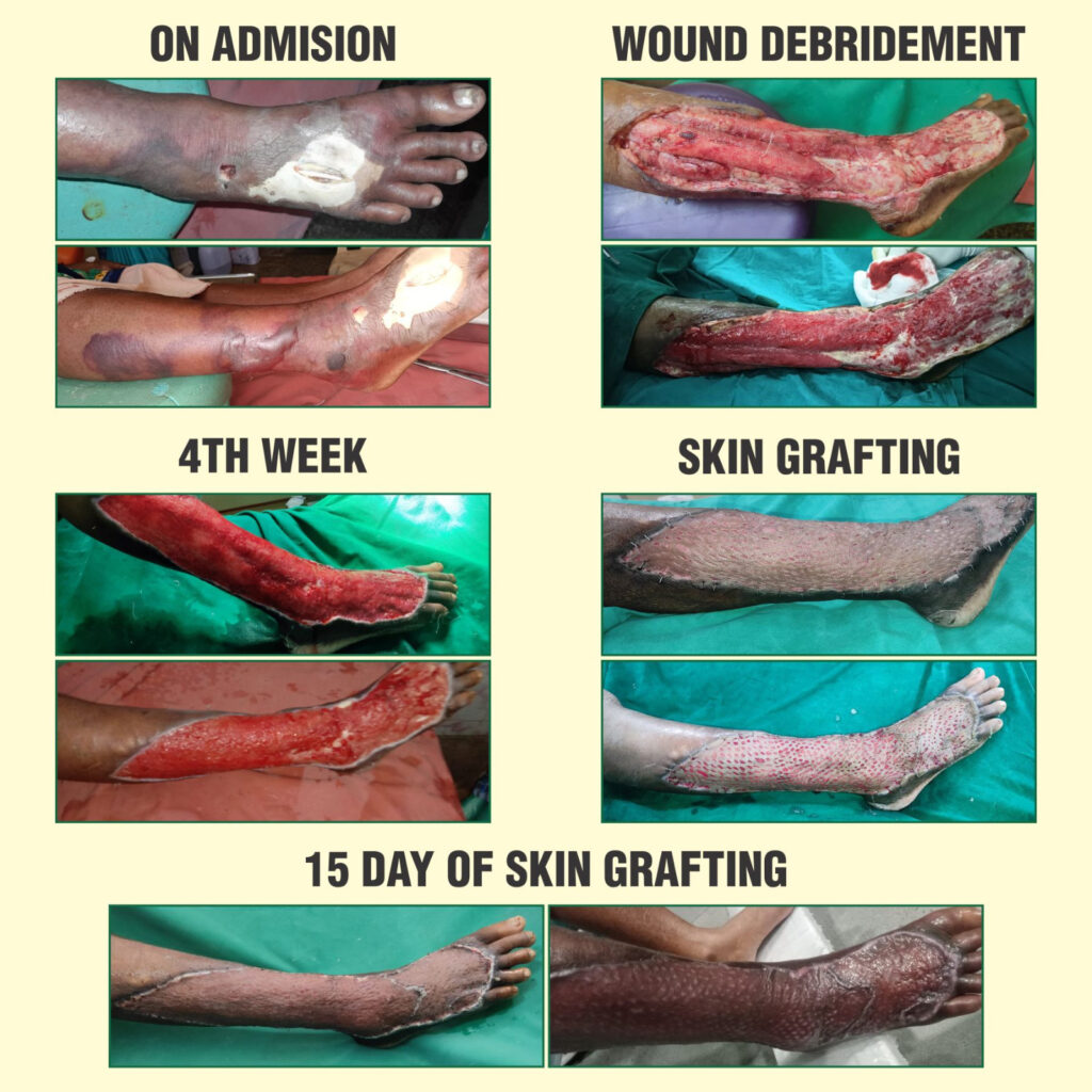
- 2வது நாள் – அறுவை சிகிச்சை செய்து பாதிக்கப்பட்ட பகுதிகளில் உள்ள அழுகிப்போன சதைகளை நீக்கினோம்.
- 15வது நாள் – நல்ல உடல்நிலை தேறியவுடன் அவரை வீட்டுக்கு அனுப்பி வைத்தோம்.
- பின்பு தினசரி வந்து ட்ரெஸ்ஸிங் போட செய்து புண் ஆறுவதற்கான முயற்சிகளை செய்தோம்.
- புண் ஆறுவதில் தாமதம் ஏற்பட்டதால் 1 மாதம் கழித்து அவரை மறுபடியும் உள் நோயாளியாக சேர்த்து தோல் ஒட்டும் அறுவை சிகிச்சை செய்யப்பட்டது.
- அறுவை சிகிச்சை முடிந்து 15 வது நாள் முழுவதுமாக அவரது புண் ஆறி நல்ல நிலையில் வீட்டுக்கு அனுப்பி வைக்கப்பட்டார்.
- தற்போது சர்க்கரை மாத்திரை சாப்பிட்டு நல்ல ஆரோக்கியத்துடன் இருக்கிறார்.
- குறிப்பு: இதற்கான ஆதார புகைப்படங்களை இணைத்துள்ளோம்.
#NECROTIZING FASCITIS
- 56 year old female patient presented with painful swelling on the Rt leg and foot for 5 days.
- Fever off and on.
- she was conscious, oriented, febrile, tachycardia.
- Local examination: Tenderness + , inflammatory signs + , pus discharge +, Multiple blisters over down of Rt foot and lower 1/3 of Rt leg.
ON EXAMINATION
HER INVESTIGATION:
- Blood Sugar: 251 mgs%
- Blood Urea: 28 mgs%
- Serum creatinine:1.1 mgs%
- HbA1c :8.8%
- Total Count: 15,000 cells/cumm [P 90%, L08%,E02%]
- Hb%: 10.8 gms%
- Urine acetone : Negative
CLINICAL DIAGNOSIS:
* Long standing T2 DM,
*Necrotizing fasciitis Rt lower limb, Involving Rt leg and Rt foot
*Septicaemia.
- She was admitted in our hospital and treated with broad spectrum antibiotics, Metronidazole, insulin infusion, iv fluids.
- After stabilization [on her 2nd admission day , She was subjected to Wound debridement done under SA.
POST OP
- Hb 8.1 gms%, 2 units of whole blood also given.
- Blood Sugar: 172 mgs%
- Blood Urea: 32 mgs%
- Serum creatinine: 1.4mgs%
- Swelling Rt leg gradually reduced in 6th post-operative day, and patient made a rapid recovery.
- Patient discharged 15th day of admission.
- Patient treated in OPD, the wound was regularly cleaned, debrided.
- The wound got red granulation tissue in 4 weeks, but not covered.
- Patient re-admitted, plan for skin grafting
- Culture and sensitivity no growth.
- Prophylactic IV Antibiotics given
- Patient already on insulin, blood sugar under control.
- Urea, creatinine normal, Hemoglobin:10.0gms%.
- Under RA, Skin grafting done by a plastic surgeon.
- Donor & recipient site healed very well (see the photo) on 15th day of Skin grafting surgery.
- This patient later on could be managed on oral antidiabetics drugs.
#NECROTIZING FASCITIS
In the tissues adjoining infections, small vessels commonly develop thrombotic occlusion leading to necrosis. After control of infections or draining, normal arterioles recanalize and form granulation tissue promoting healing of the wound. However, in certain mixed infections, so common in diabetic foot, virulent strains of staphylococci, and streptococci liberate angiotoxic (necrotizing) substance.
These angiotoxic factors like alpha-toxin of staphylococci, along with spreading factors like streptokinase and streptococcal hyaluronidase lead to rapid extension of necrosis by digestion of fibrin barriers and intracellular ground substance, this is severe, progressive soft tissues infection, which leads to necrosis of subcutaneous tissues down to the level of muscle fascia.
Clinically, it begins with a trivial infection in the foot or hand leading to rapidly advancing necrosis with erythema, swelling, and dusky discoloration of the overlying skin with haemorrhagic blisters.
The occlusive process in the small vessels is exaggerated more arterioles are obliterated and the original lesions get converted from trivial to ever enlarging areas
of necrosis and gangrene.Creeping advancement of this process of infective obliterative angiopathy leads to devasting lesions within a couple of days.
There is also evidence of systemic toxicity in the form of fever, tachycardia, and hypotension.Urgent fasciotomy, fascietomy and if required a major amuputation needs to be carried out of along with an aggressive medical treatment to prevent such a life threatening situation.
References:
DIABETIC FOOT:
A Clinical Atlas / Sharad Pendsey