- 67 year old female patient presented with painful swelling on the Rt foot for 5 days.
- Fever off and on.
- ON EXAMINATION
- she was conscious, oriented, febrile, tachycardia.
- Local examination: Tenderness + , inflammatory signs + , pus discharge + over the 1st MTP region.
HER INVESTIGATION:
- Blood Sugar: 318 mgs%
- Blood Urea: 46 mgs%
- Serum creatinine:1.5 mgs%
- HbA1c :8.5%
- Total Count: 14,900 cells/cumm [P 90%, L08%,E02%]
- Hb%: 8.1 gms%
- Urine acetone : Negative
CLINICAL DIAGNOSIS:
* Gangrene right great toe with cellulitis right foot
* AKI
* Septicaemia,
* Anaemia.
- She was admitted in our hospital and treated with broad spectrum antibiotics, Metronidazole, insulin infusion, iv fluids.
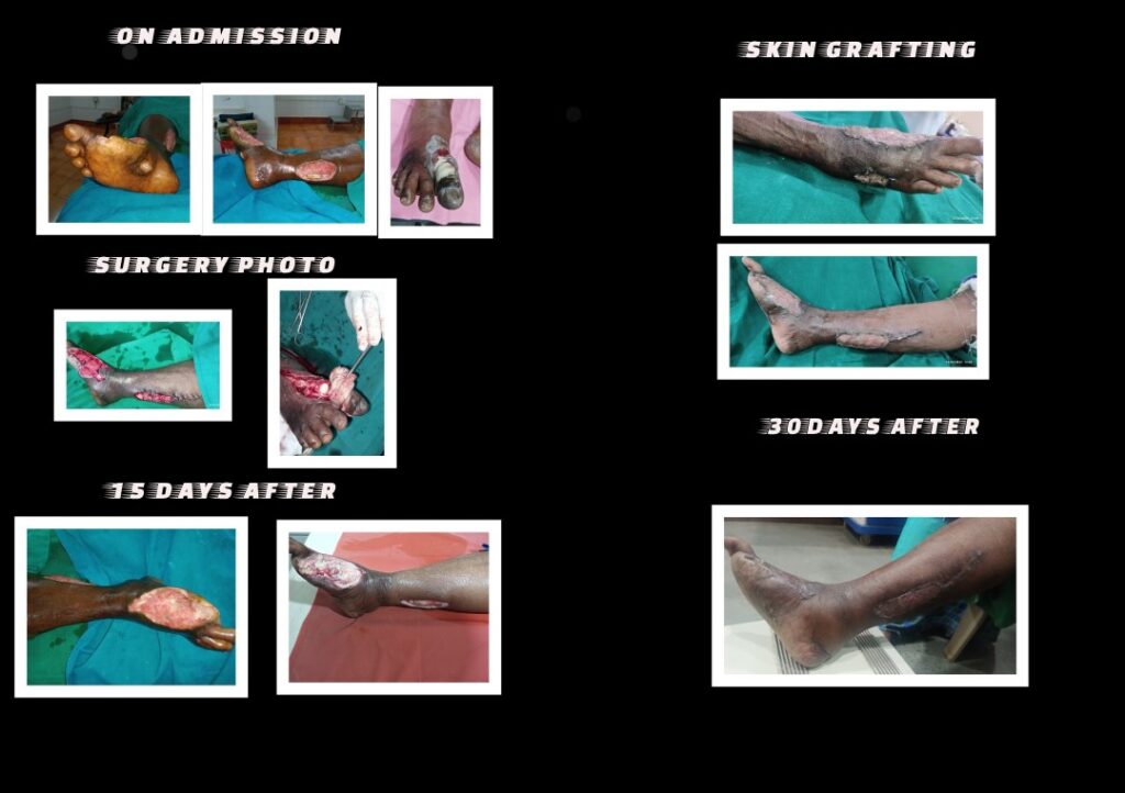
- After stabilization [on her 2nd admission day , She was subjected to Ray amputation of right great toe and fasciotomy done under RA.
POST OP
- 1unit of whole blood, 1Unit of PCV also given.
- Blood Sugar: 178 mgs%
- Hb 9.5 gms%,
- Blood Urea: 32 mgs%
- Serum creatinine: 1.4mgs%
- Swelling Rt leg gradually reduced in 5th post-operative day, and patient made a rapid recovery.
- Patient discharged 8th day of admission.
- Patient treated in OPD, the wound was regularly cleaned, debrided.
- The wound got red granulation tissue in 3 weeks, but not covered.
- Patient re-admitted, plan for skin grafting
Culture report: Acinetobacter baumanii grown in culture.
Antibiotics given asper culture report.
- Patient already on insulin, blood sugar under control.
- Urea, creatinine normal, Hemoglobin:10.0gms%.
- Under SA, Skin grafting done by a plastic surgeon.
- Donor & recipient site healed very well (see the photo) on 13th day of Skin grafting surgery.
- This patient later on could be managed on oral antidiabetics drugs.
#GANGRENE
WET GANGRENE:
The tissues are gray or black, moist and often malodorus, affected part is swollen and the epidermis may be raised in blebs, adjoining tissues are infected and pus may discharge from ulcerated areas.
References:
DIABETIC FOOT: 2003 Page no: 127
A Clinical Atlas / Sharad Pendsey