TAMIL VERSION:
92 வயதான அம்மா.
- கடந்த 22 ஆண்டுகளாக எங்கள் மருத்துவமனையில் சர்க்கரை நோய்க்கு முறையான சிகிச்சை எடுத்துக் கொண்டிருக்கிறார்.
- ஒரு சிறிய விபத்தில் இடது முழங்காலில் அடிபட்டு விட்டது.
- இடது முழங்காலில் வீக்கம் ஏற்பட்டு வலி அதிகம் இருந்தது.
- அவரை மருத்துவமனையில் அட்மிஷன் செய்தோம்.
- முறையாக வைத்தியம் செய்ததால் அவரது ரத்த சர்க்கரைஅளவு நல்ல கட்டுப்பாட்டில் இருந்தது.
- சர்க்கரை அளவு 182 mgs%
- மூன்று மாத சர்க்கரை அளவு HbA1c 7.8%
- அவரது கிட்னி செயல்பாடும் நன்றாக இருந்தது.
- அவருக்கு முறையான ஆண்டிபயாட்டிக் மருந்துகள் கொடுத்தோம்.
- சர்க்கரையை இன்னும் கட்டுப்படுத்த இன்சுலின் ஊசி பயன்படுத்தினோம்.
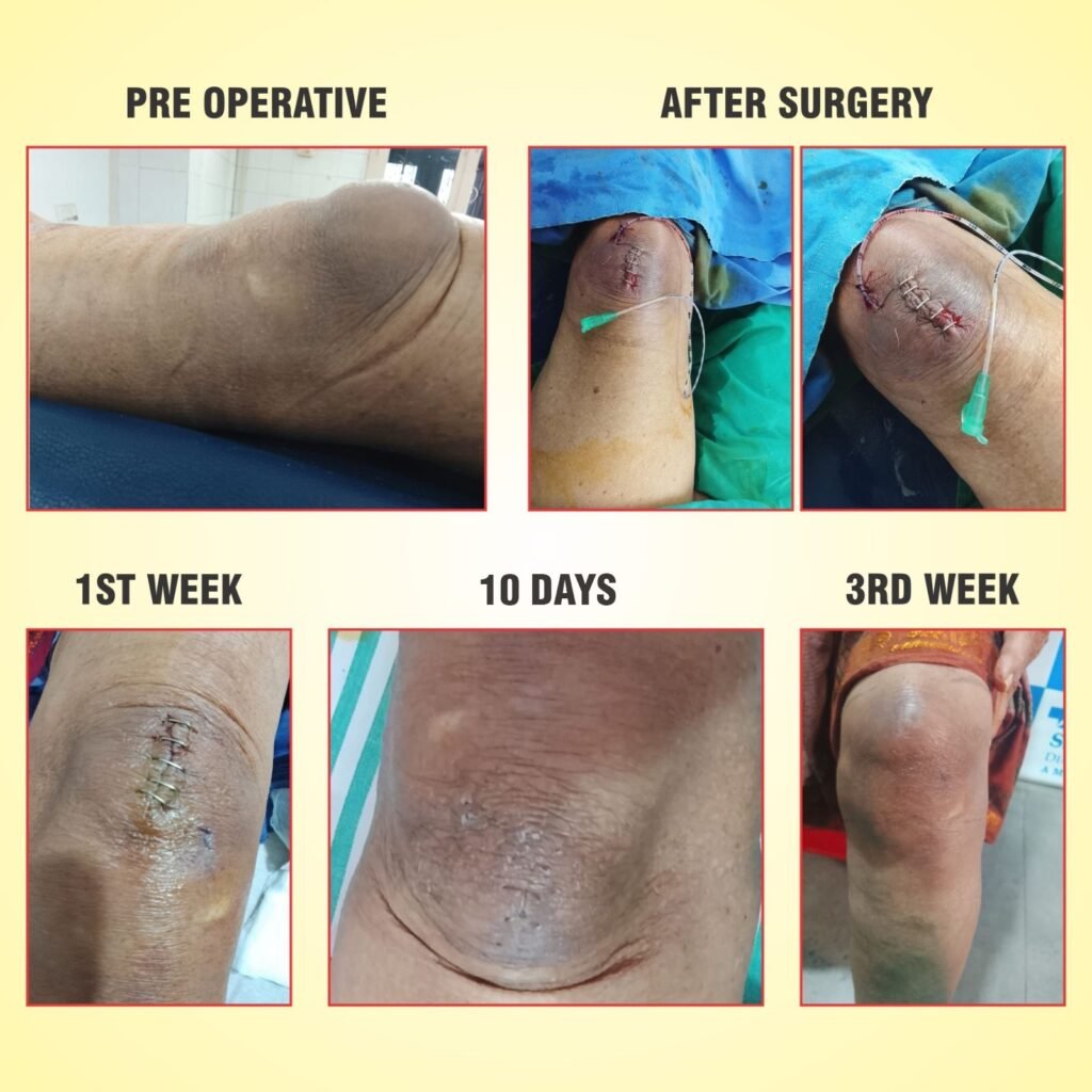
- 2வது நாள் அவரது காலில் ஒரு சிறிய அறுவை சிகிச்சை செய்து காலில் உள்ள உறைந்து போன ரத்தம் பாதிக்கப்பட்ட பகுதிகளை நீக்கி விட்டோம்.
- 4வது நாள் நல்ல முன்னேற்றம் ஏற்படவே அவரை வீட்டுக்கு அனுப்பி வைத்தோம்.
- அதற்குப் பின் அவர் தொடர்ச்சியாக வந்து கால் புண்ணிற்கு ட்ரெஸ்ஸிங் செய்து கொண்டார்.
- 10 நாட்களில் அவரது புண் முழுவதுமாக ஆறிவிட்டது.
- அதன் பின் இன்சுலின் நிறுத்திவிட்டு மாத்திரை போட்டுக் கொண்டு நலமுடன் வாழ்ந்து வருகிறார்.
குறிப்பு:
இது சிறிய அறுவை சிகிச்சை தான், இருப்பினும் அவருடைய வயது 92.
சர்க்கரை கட்டுப்பாட்டில் இருந்தால் எந்த வயதிலும் அறுவை சிகிச்சை செய்ய முடியும் என்பதற்கு இவரே சாட்சி.
இதற்கான படங்களை பதிவிட்டுள்ளோம்.
நன்றி
ENGLISH VERSION:
A 92-year-old female patient presented with pain and swelling over the left knee for 10 days following a history of fall and injury to the left knee.
She had been on regular treatment for Type 2 Diabetes Mellitus at our hospital for the past 22 years.
Clinical Examination
The patient was conscious and oriented.
Local Examination – (Left Knee)
Tenderness and Inflammatory signs present over the swelling in the left knee region.
Investigations
Random Blood Sugar: 182mg/dL
Blood Urea: 29 mg/dL
Serum Creatinine: 1.1 mg/dL
HbA1c: 7.8%
Total Count: 6,800 cells/cumm
Hemoglobin: 10.8 g/dL
Clinical Diagnosis
Type 2 Diabetes Mellitus
Left knee pre patellar hematoma following trauma
Treatment Given
The patient was admitted and started on Broad-spectrum antibiotics, Metronidazole and
Insulin therapy
2nd day of admission, the patient underwent incision and drainage (I&D) of the left knee hematoma.
Post-Operative Course
Post-operative investigations revealed:
Postprandial Blood Sugar: 103 mg/dL
Hemoglobin: 10 g/dL
Blood Urea: 26 mg/dL
Serum Creatinine: 1.0 mg/dL
The swelling gradually reduced by the She 3rd post-operative day, and the patient showed significant clinical improvement. She made a rapid recovery and was discharged on the 4th day of admission.
Follow-Up
Regular wound cleaning and dressing were performed.
Oral Antibiotics were continued.
Blood sugar levels remained under control with insulin therapy.
Renal parameters remained within normal limits.
Hemoglobin stabilized at 10 g/dL.
Wound healed well, suture remove after 10 days.
Subsequently, the patient was successfully managed with oral antidiabetic medications.
She has been on regular treatment at our hospital for the past 22 years, and her diabetes has been well controlled. As a result, she had no major complications.
Even at the age of 92, the procedure was done successfully, and she recovered well and went home after four days.